Diagnostics
- Duplex sonography (ultrasound diagnostics)
- Electroneurography/electromyography
- Electroencephalography (EEG)
- Evoked potentials
- Tests for memory disorders
Ultrasound diagnostics / duplex sonography
This ultrasound method allows the blood flow in the main arteries in the neck and head to be visualised. We can use this examination to detect vasoconstrictions and vascular calcifications and assess their severity. Vascular constrictions are a frequent cause of strokes.
The examination takes approx. 15-20 minutes.
In individual cases, this duplex sonography is also one of the IGeL services.
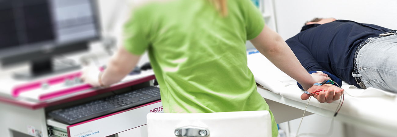
Electroneurography / electromyography
Electroneurography, ENG
In electroneurography, a nerve is stimulated using a brief electrical stimulus. This allows the nerve conduction velocity and amplitude, the number of nerve fibres stimulated, to be determined.
This method can be used to examine clinical pictures such as carpal tunnel syndrome, nerve injuries or polyneuropathies. The examination takes between 10 and 30 minutes, depending on the problem.
Electromyography, EMG
Electromyography uses a thin needle to measure electrical muscle activity in muscles. This method can be used to differentiate between various muscle and nerve diseases. Acute and chronic changes can also be differentiated.
Electroneurography and electromyography are often combined.
Electroencephalography (EEG)
With the help of the EEG, the electrical activity of the brain can be determined by recording the voltage fluctuations on the surface of the brain. Electrodes are attached to various points on the head.
This examination is used to clarify and investigate the course of epileptic disorders.
An EEG examination takes about 20 minutes. The electrodes are placed on the scalp using a cap.
Evoked potentials
Evoked potentials are specifically triggered bioelectric potentials to assess the visual and auditory pathways and the sensory pathways. These examinations take about 10 minutes each.
Visual evoked potentials (VEP)
In the VEP examination, the visual pathway/ auditory cortex is optically stimulated using a black and white chequerboard pattern. It is used, for example, to examine acute and chronic inflammation of the visual pathway.
Acoustic evoked potentials (AEP)
In the AEP examination, the auditory pathway is stimulated by side-separated stimulation with clicking noises via headphones. This examination can help to clarify dizziness and also inflammatory diseases of the central nervous system.
Somatosensory evoked potentials (SSEP)
Sensory nerve pathways are stimulated by stimulating individual nerves in the arms and legs. SSEP is carried out in the case of unexplained sensory symptoms caused by a disorder of the central nervous system, in particular the spinal cord.
Tests for memory disorders
We carry out various tests to clarify a memory disorder.
The choice of tests depends on the underlying disease or suspected diagnosis.
We offer the following tests:
MMST, DemTect, clock test, Panda, MOCA, SKT, FSMC.
Kontakt
Tel. allgemein:
030 | 930 93 56
Tel. Privatpatient:innen:
030 | 935 238 69
FAX: 030 | 284 769 04